当前位置:网站首页>The application of AI in the whole process of medical imaging equipment
The application of AI in the whole process of medical imaging equipment
2022-07-31 02:11:00 【IT Geek Gang】
AI is no longer a commercial gimmick. It has applications in various industries. Today, let's take a look at how AI can accompany and help the development of imaging equipment in the medical industry.
AI mainly solves three problems. First, AI can be used as an experienced doctor to improve the diagnosis and treatment level of grassroots hospitals. Second, AI can be used as an assistant to relieve doctors from complicated work. Third,Explore patterns in big data.
Let's reveal the role of AI from the entire workflow of imaging equipment:
(1) Scanning Stage
Smart Positioning:
The positioning work is mainly completed by the operating technician, who completes the preliminary positioning according to the scanning protocol. This method has low operation efficiency and poses a radiation safety hazard to the scanning technician. In view of this, AI can be used to complete the automatic positioning process.With the help of the high-definition camera installed above the device, the patient's photos are taken, the anatomical key points are automatically identified, and the bed movement size is automatically calculated according to the camera calibration parameters and scanning protocol.

Automatically position images:
The positioning process is mainly based on the low-dose positioning image to accurately set the scanning range and angle. Compared with CT, MR can scan at any angle, so the angle information is very important. For example, by dividing the headSagittal image of the corpus callosum, with the rotation of the bounding box angle through the corpus callosum as the scan angle.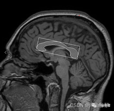
Image reconstruction:
After the scanning is completed, the CT detector receives the X-ray attenuation signal after passing through the human body, and the MR receiving coil receives the tissue hydrogen proton relaxation and releases the energy signal, which is converted into digital signal by analog-to-digital conversion. The reconstruction process is to convert these digital signals into humanAn understandable grayscale image.
CT image quality is related to radiation dose, and MR image quality is related to scan time. The reconstruction algorithm is to solve how to obtain high-quality images at low dose and in a short time.
Reconstruction algorithms can be divided into: algebraic reconstruction, filtered back-projection, iterative reconstruction, and deep learning reconstruction according to their development history.
There are also two types of reconstruction algorithms based on deep learning. One uses raw raw data as input and outputs high-quality reconstructed grayscale images. Typical examples are GE's Turefidelity and Canon's Aice.

Turefidelity is taken as an example. Its training data uses two scanning parameters to obtain high-quality and low-quality images respectively. The model takes the low-quality image as input, and compares the predicted output image with the scanned high-quality image to calculate the loss function to optimize the parameters..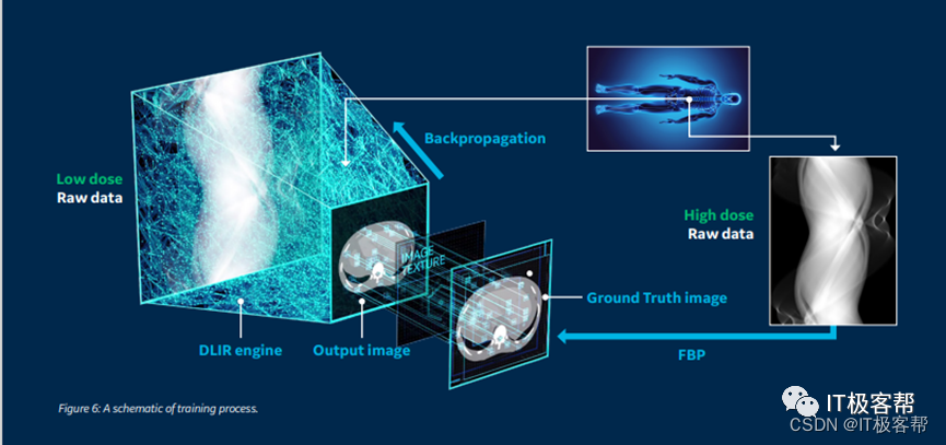
The other is the reconstructed grayscaleThe image is used as input to perform operations such as denoising, de-artifacting, super-resolution, etc.
(2) Stage of diagnosis and treatment:
There are countless applications of AI in the diagnosis and treatment stage. According to the application in the field of vision, it can be divided into image classification, target detection, image segmentation, image generation, etc.
Image classification: As a qualitative analysis method, it can provide doctors with a basis for diagnosis, diagnosis of benign and malignant tumors, prediction of bone age, rapid classification of stroke, etc.
Object detection: As a semi-quantitative analysis method, it can not only provide classification results, but also provide the location and size of ROI, such as lung nodule detection.
Image segmentation: As a full quantitative analysis method, it can provide pixel-level classification results, and then calculate ROI volume, maximum diameter and other parameters, such as tumor segmentation, organ segmentation, etc.
Image generation: It mainly solves the problem of insufficient medical image data.
边栏推荐
猜你喜欢
mmdetection trains a model related command

leetcode-1161: Maximum in-layer element sum

Fiddler captures packets to simulate weak network environment testing
The effective square of the test (one question of the day 7/29)

Teach you how to configure Jenkins automated email notifications
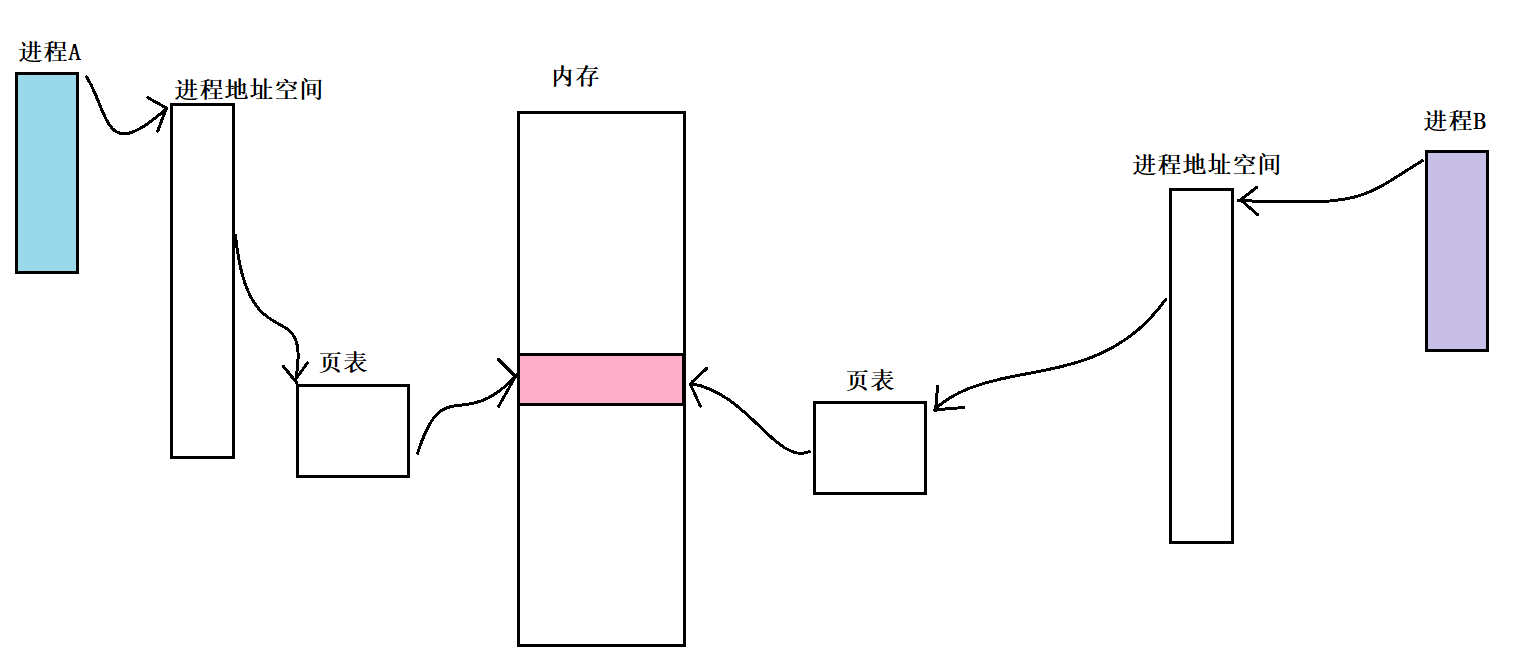
Interprocess communication study notes
最高月薪20K?平均薪资近万...在华为子公司工作是什么体验?

CV-Model【3】:MobileNet v2
![[Map and Set] LeetCode & Niu Ke exercise](/img/66/d812a6ad854cb0993c796760042150.png)
[Map and Set] LeetCode & Niu Ke exercise

Crypto Life, a day in the life of a Web3 project partner
随机推荐
Verify the integer input
Basic introduction to ShardingJDBC
mysql index
221. Largest Square
Force buckled brush the stairs (7/30)
用户交互+格式化输出
真正的CTO,是一个懂产品的技术人
coldfusion文件读取漏洞(CVE-2010-2861)
To write good test cases, you must first learn test design
C language applet -- common classic practice questions
CV-Model [3]: MobileNet v2
Detailed explanation of STP election (step + case)
基于FPGA的售货机
leetcode-399:除法求值
multiplayer-hlap 包有问题,无法升级的解决方案
Introduction and use of Drools WorkBench
How to expose Prometheus metrics in go programs
Crypto Life, a day in the life of a Web3 project partner
Arbitrum 专访 | L2 Summer, 脱颖而出的 Arbitrum 为开发者带来了什么?
Gateway routing configuration