当前位置:网站首页>FFA and ICGA angiography
FFA and ICGA angiography
2022-07-07 23:46:00 【Microelectronics and solid state electronics - Yu Chi】
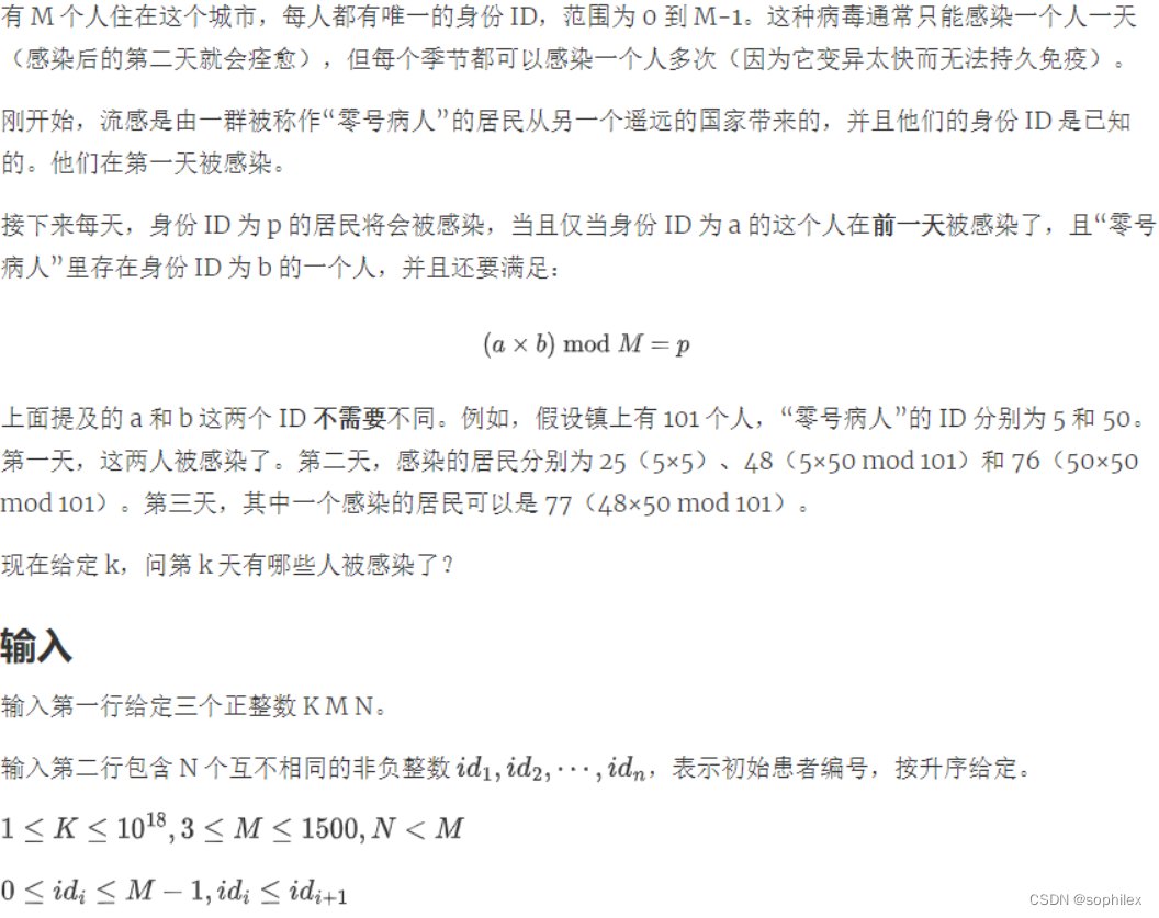
| Type of angiography | Contrast object | Anatomy | Structure Vascular proliferation |
| FFA( Sodium fluorescein ) | Retinal vessels | The fundus oculi | 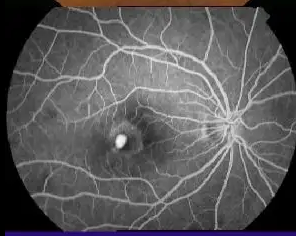 |
| ICGA( Indocyanine green ) | Choroid | Extrabulbar | 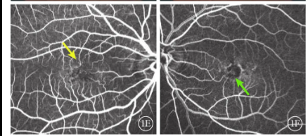 |
Some patients go to experts , The expert replied that there was no hyperplasia , Let's keep observing 、 Follow up regularly .
Then the patient went back happily ,
What's going on here ?
This is because fluorescein angiography shows no leakage , If there is penetration ( It is most likely caused by neovascularization ) It's time to damage the photosensitive layer .
So at this point , It's best to control yourself in advance ( For example, anti vegf Relevant diet, etc )
#######################################################################

chart 1: Left for FFA Images , It can be seen that there is only a small amount of fluorescent leakage in the macular area , The lack of CNV Typical performance ; The right to ICGA Images , It can clearly show pigment epithelium detachment and CNV ingredients , Provide richer information for disease diagnosis .
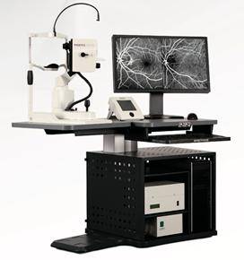
chart 2: Heidelberg HRA

chart 3: wAMD In patients with FFA+ICGA Early image of synchronous angiography

chart 4: wAMD In patients with FFA+ICGA Synchronous late imaging

chart 5: FFA+ICGA Early display of synchronous angiography PCV Abnormal branch vascular network and polypoid lesions

chart 6: Another patient ICGA+OCT:ICGA The location of polypoid lesion in OCT There are many pigment epithelium detachment (PED),PED Incisure ( White arrow ) And for polyps PED peak ( Inverted triangle ) It can be called “ Thumb sign ”[2].
边栏推荐
- P2141 [noip2014 popularization group] abacus mental arithmetic test
- C simple question one
- One of the anti climbing methods
- C method question 1
- Archery installation test
- Chisel tutorial - 02 Chisel environment configuration and implementation and testing of the first chisel module
- May day C - most
- FFA与ICGA造影
- SAP HR family member information
- Pycharm essential plug-in, change the background (self use, continuous update) | CSDN creation punch in
猜你喜欢
![[STM32 + esp-12s connect Tencent cloud IOT development platform 1] creation of cloud platform and burning of at firmware](/img/bc/8241a339cca9b7af475169dba39c10.jpg)
[STM32 + esp-12s connect Tencent cloud IOT development platform 1] creation of cloud platform and burning of at firmware
SAP HR family member information
神奇快速幂
关于CH32库函数与STM32库函数的区别

一份假Offer如何盗走了「Axie infinity」5.4亿美元?
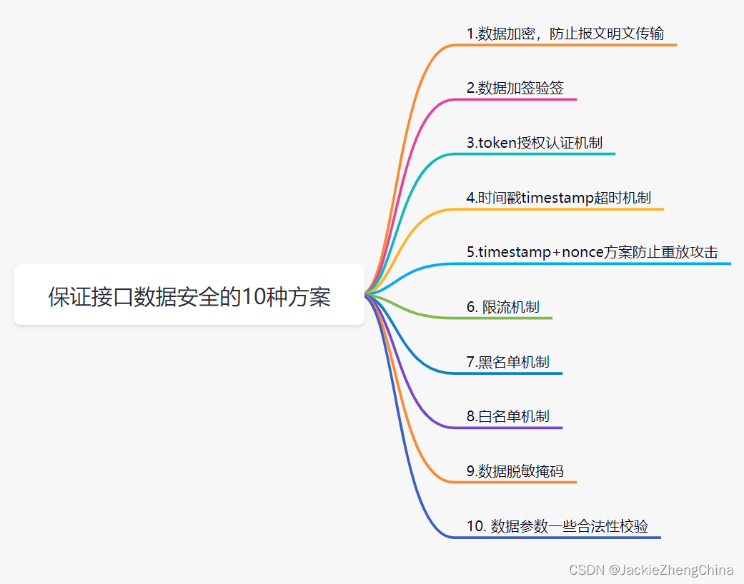
10 schemes to ensure interface data security
![[stm32+esp8266 connects to Tencent cloud IOT development platform 3] stm32+esp8266-01s dynamically registers devices on Tencent cloud (at instruction mode) -- with source code](/img/55/ab50ead2564498cb214d98ac5b9c3d.jpg)
[stm32+esp8266 connects to Tencent cloud IOT development platform 3] stm32+esp8266-01s dynamically registers devices on Tencent cloud (at instruction mode) -- with source code
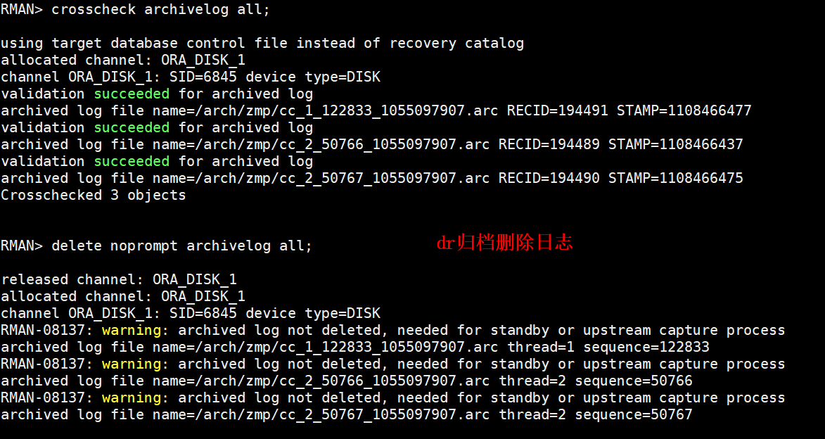
DataGuard active / standby cleanup archive settings

Chisel tutorial - 04 Control flow in chisel
Svn relocation
随机推荐
C cat and dog
aws-aws help报错
Anti climbing means cracking the second
Slam interview summary
Display the server hard disk image to the browser through Servlet
JNI uses asan to check memory leaks
C # exchange number, judge to pass the exam
保证接口数据安全的10种方案
C - linear table
Archery installation test
archery安装测试
Take you hand in hand to build feign with idea
SAP HR labor contract information 0016
Enterprise application demand-oriented development of human resources department, employee attendance records and paid wages business process cases
数据分析系列 之3σ规则/依据拉依达准则来剔除异常值
Chisel tutorial - 05 Sequential logic in chisel (including explicit multi clock, explicit synchronous reset and explicit asynchronous reset)
P1055 [noip2008 popularization group] ISBN number
MongoDB快速入门
Go time package common functions
redis缓存工具类,值得拥有~